Learning how to see in three dimensions while looking at an image that is only showing you two dimensions is an art that takes time to accomplish. Sure there is equipment out there that scans in three dimensions, and you will probably encounter them during your education, but in reality almost all of your scanning will still be performed utilizing 2-D technology. That does not mean you can forget about that third dimension. It is always there; you just have to learn how to “see” it.
To help with this, I like to draw. Yes, drawing is done in two dimensions as well. But if you draw two images of the same structure at ninety degrees from each other, then draw a pie shape representing your transducer and beam, you will begin to see what I mean. You can also move the pie shape around the structure to see what you will be looking at from different windows and angles. Your drawings can be very simple, stick figure ultrasounds if you will. You do not have to be an artist here. Just draw the basic outline of the structure. For instance here is a drawing of the liver in longitudinal orientation:
 Longitudinal

And a drawing of a liver in transverse orientation:
 Transverse
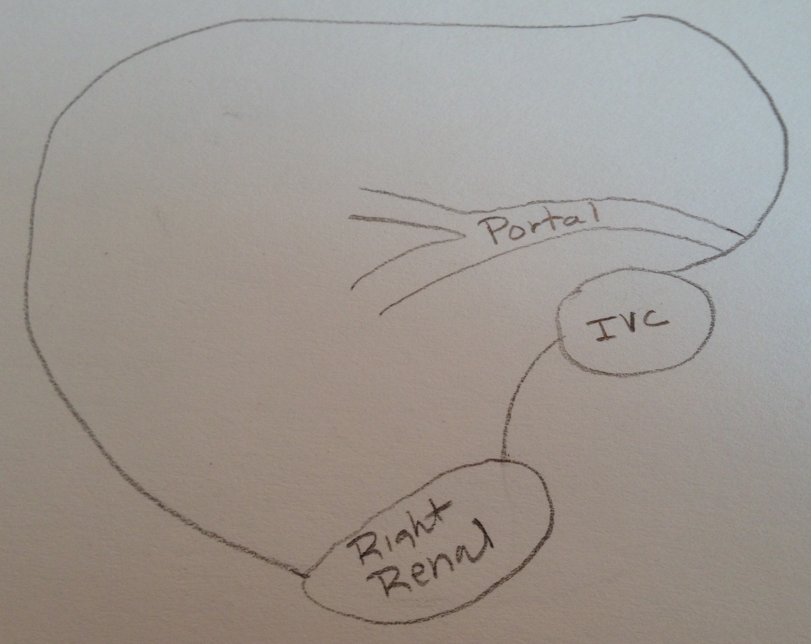
It is useful to place something inside the structure, where a vessel or mass may be. If you place the transducer on the anterior surface of the abdomen and simply turn it from longitudinal to transverse, these are the images you will see:

 Longitudinal
 Transverse
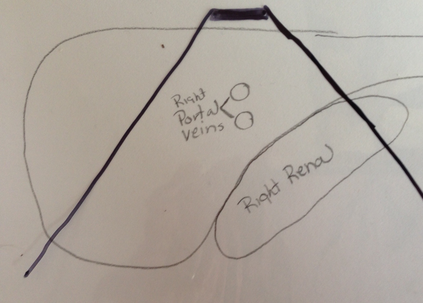
But what happens when you move the transducer to a lateral window? Using the transverse image this is what it would look like:
 Transverse Coronal
 The lateral edge of the liver now is what is closest to the transducer face, so instead of looking at the liver in an anterior/posterior (AP) slice, you will now be seeing it in a coronal slice. If you then turn the transducer back to a longitudinal orientation at the level of the IVC this is what you will see:
 Longitudinal Coronal
It may not look too much different when just looking at a normal liver, but will be tremendously important when trying to localize something or simply trying to visualize in your head the whole organ while only looking at a slice of it.
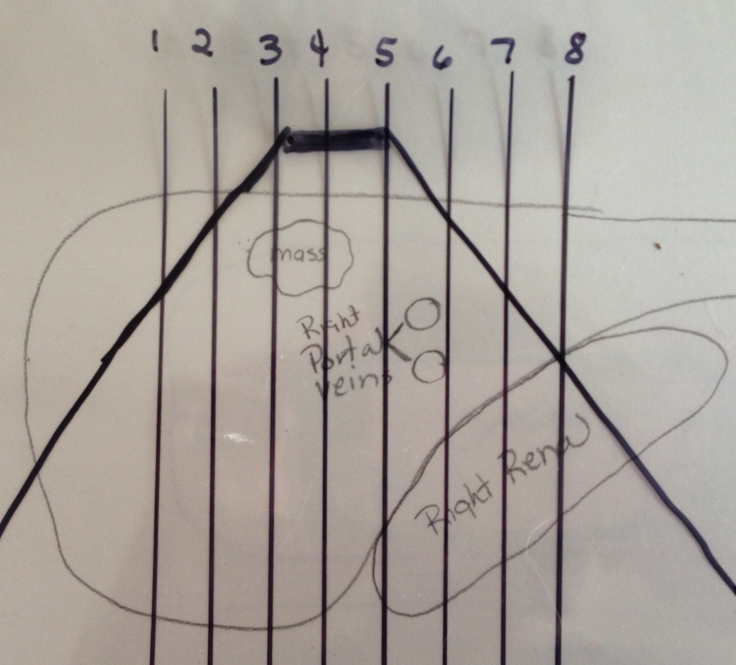
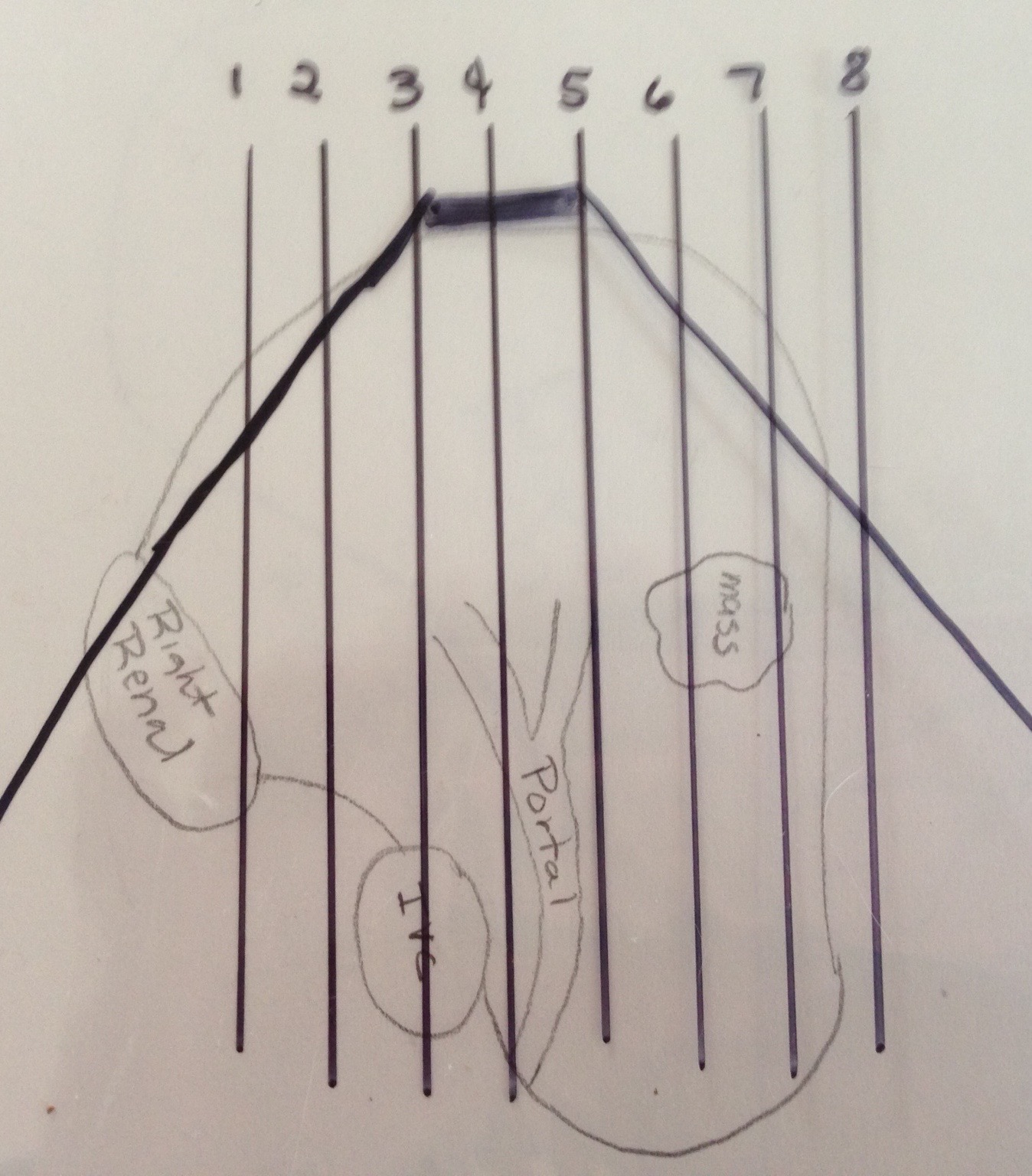
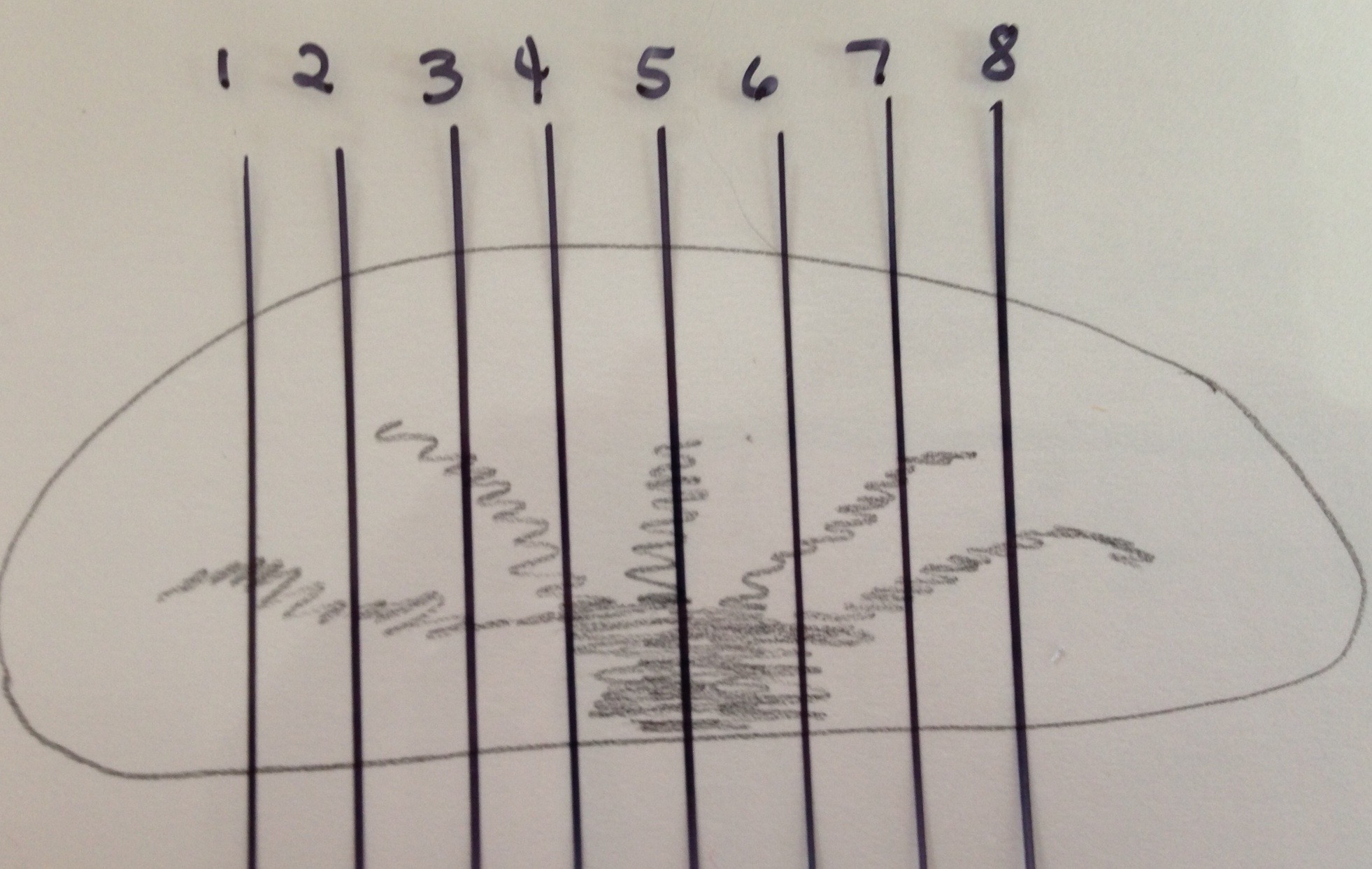
So now place a mass into that liver. First look at it in a longitudinal orientation:
 Longitudinal
Drawing vertical plane lines throughout will help you to understand where you should see the lesion when scanning transverse. When then turning transverse you can move to the correct slice to see it. This next image would be transverse at the level of the third or fourth line:
Transverse
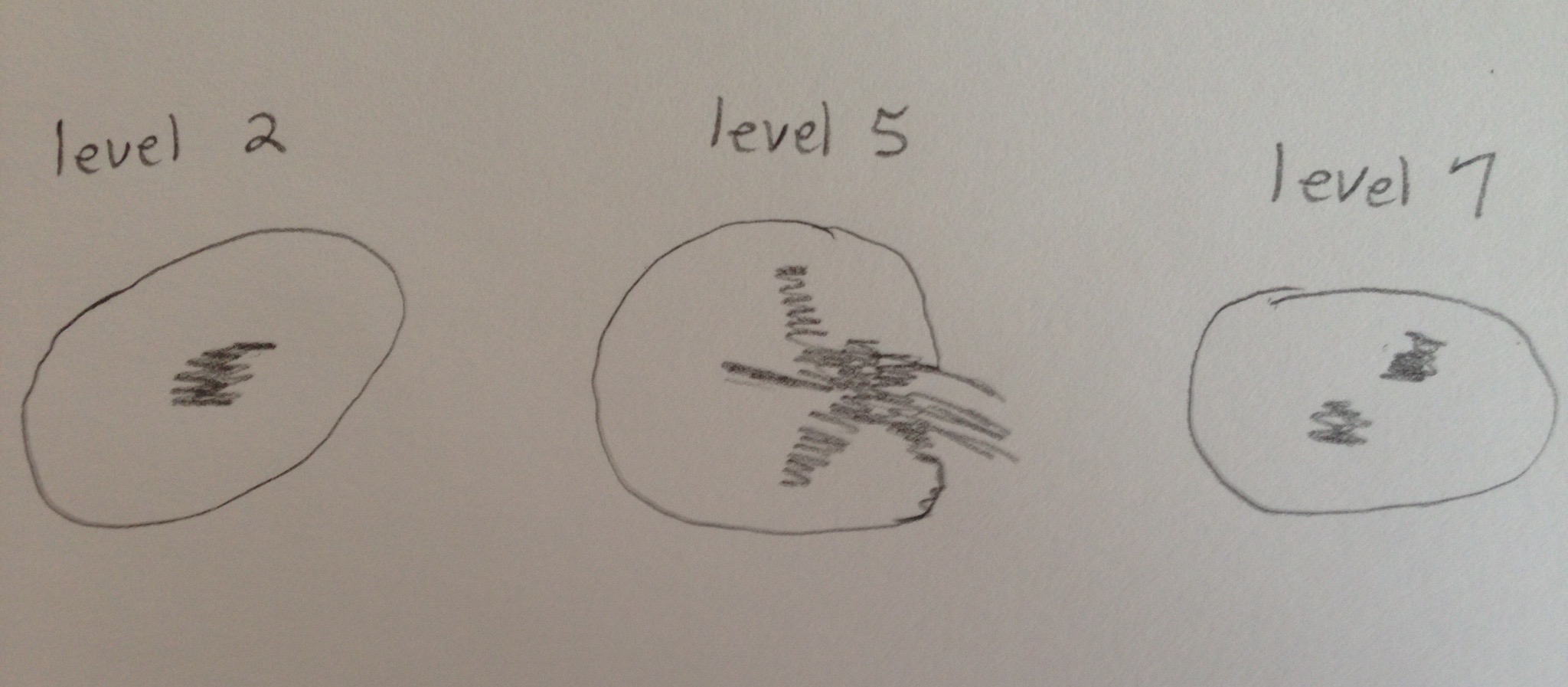
But you can also see where it would be if we moved the transducer to a different window:
 Transverse Coronal
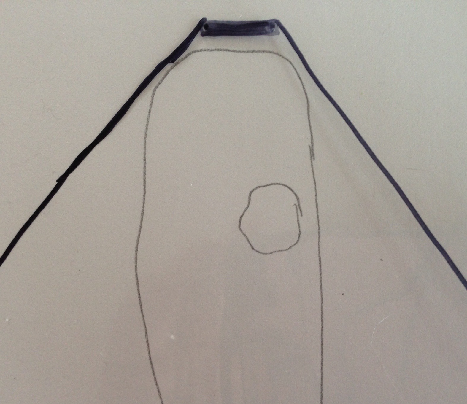
And then while using that window you could again use lines to determine what slice you would see it at in a longitudinal plane:

 Transverse Coronal
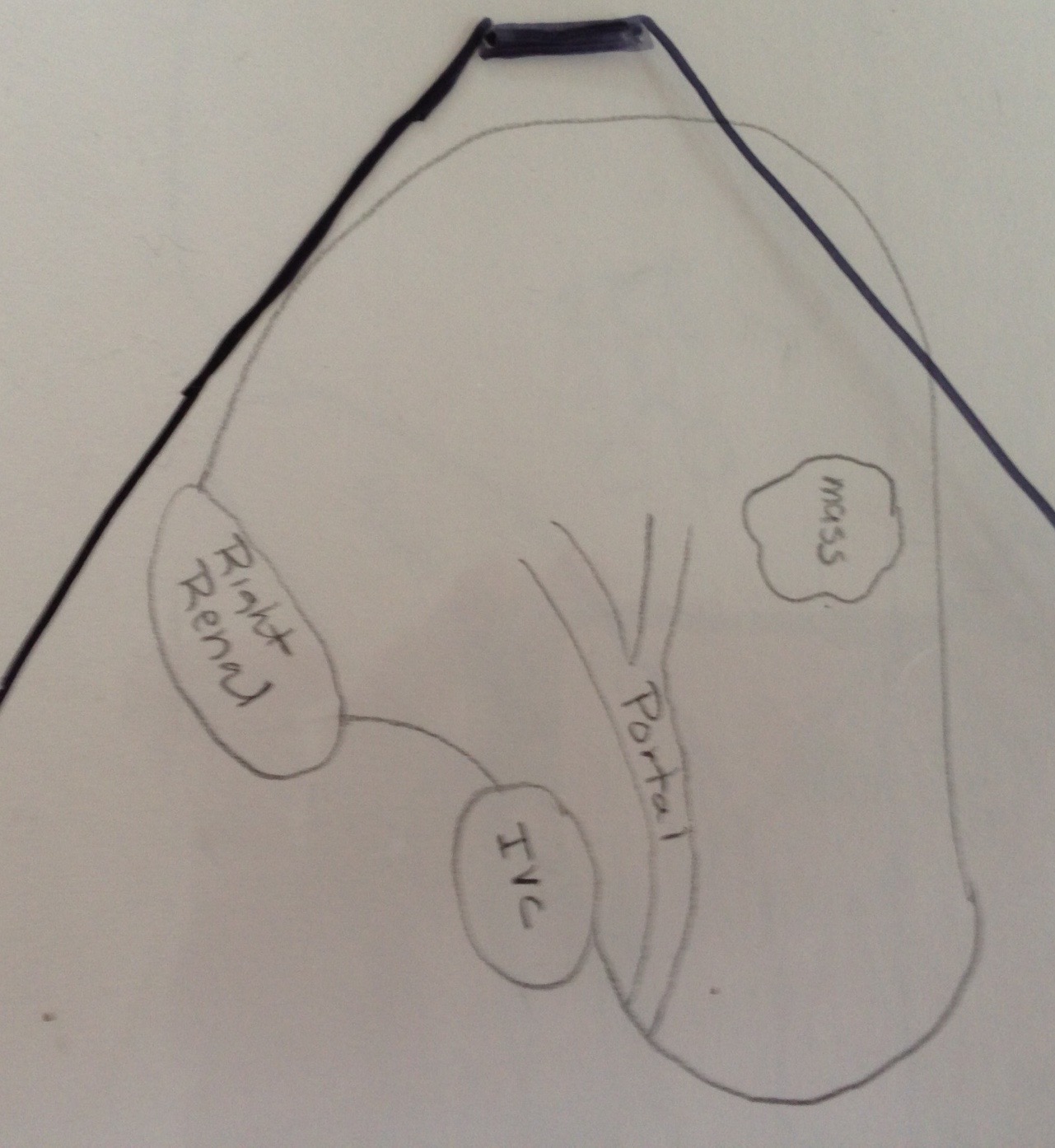
In this case, we would best see it at the level of line six. Because we are so far up towards the anterior surface of the liver, we would no longer see the IVC or right portal vein in the longitudinal plane:
 Longitudinal Coronal
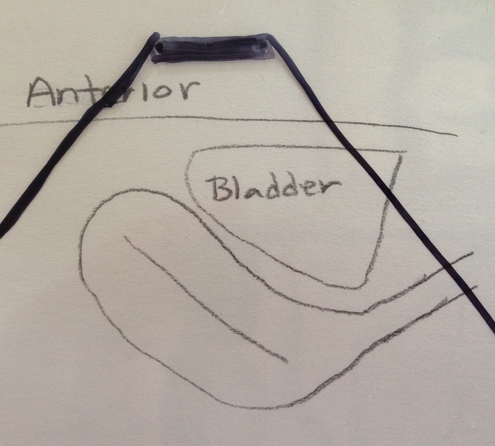
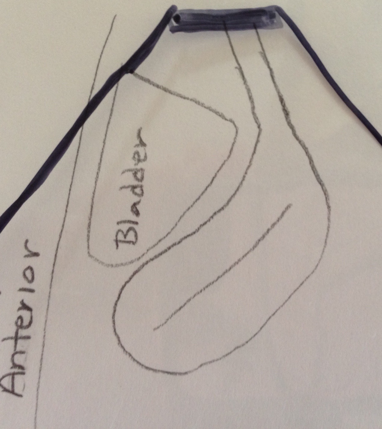
This technique is especially helpful when learning to scan endo-vaginally to get your orientation of what is anterior and posterior:
 Trans-abdominal anteverted uterus

 Endo-vaginal anteverted uterus

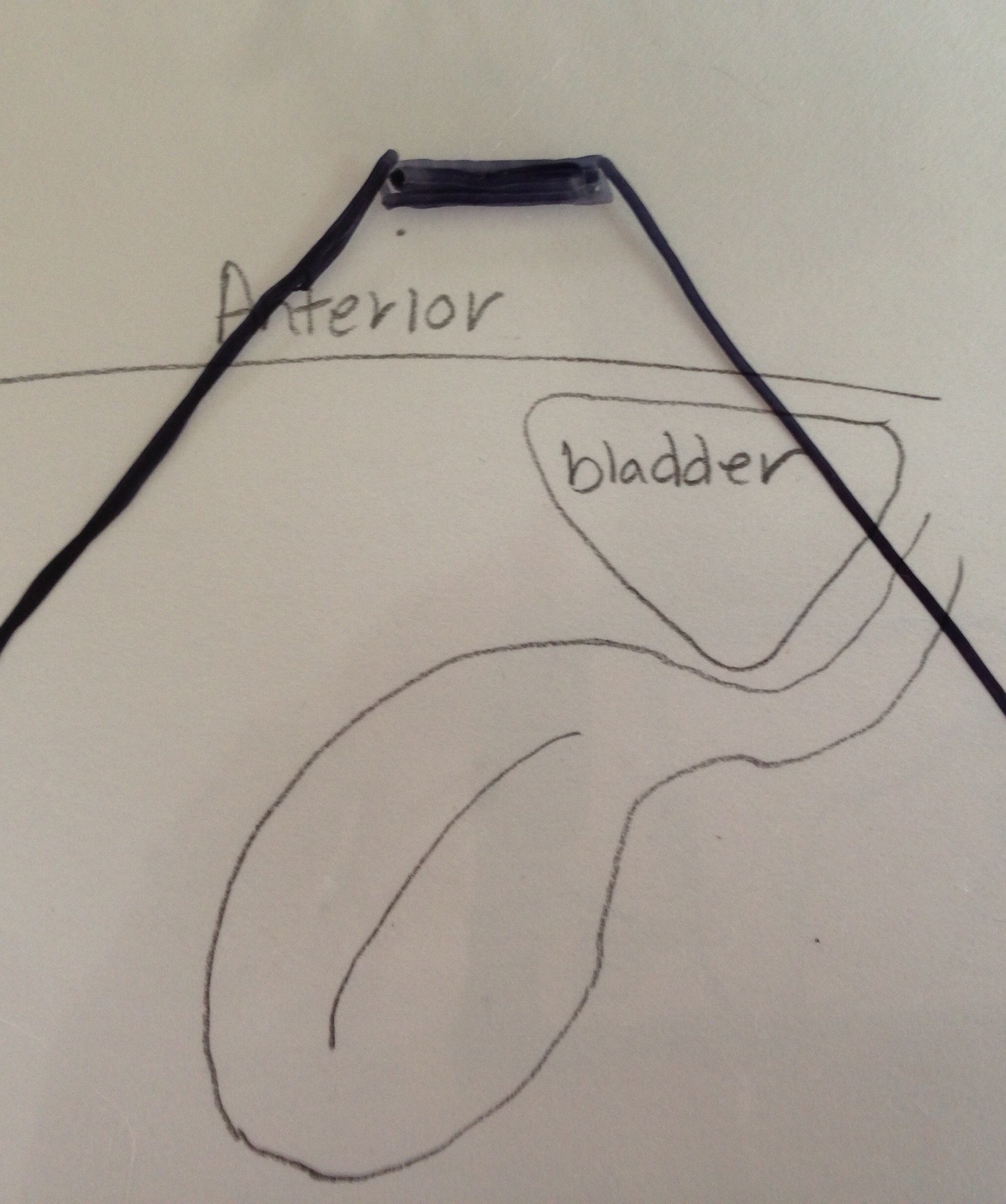 Trans-abdominal retroverted uterus
 Endo-vaginal retroverted uterus
It can also be useful to take one organ, such as a kidney, and draw a longitudinal image with several vertical cut lines through it, then drawing the transverse representation of each of those lines.
 Longitudinal
 Transverse

Use this technique for any organ you are having trouble visualizing in your head in three dimensions. It should greatly help you “see” it in your mind and, therefore, understand what you see on the image in front of you and where to move your transducer to see it from any window. It can also help you understand what happens when you physically move your transducer versus when you angle your transducer from a stationary point. Pretty soon you should be able to visualize without the drawing.